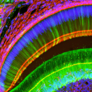
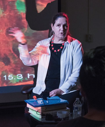
SARAH DALLAS, PH.D., doesn’t just look at cells through a microscope. The director of UMKC’s Confocal Microscopy Core uses her high-powered devices to actually watch living cells at work.
Dallas is a University of Missouri Curator’s Distinguished Professor and serves as the Lee M. and William Lefkowitz Endowed Professor in the School of Dentistry’s Department of Oral and Craniofacial Sciences. She recently received a $600,000 National Institutes of Health shared instrumentation program grant to purchase a new confocal microscope that will provide her an even better view of cell life.
The devices Dallas uses and offers to the entire community of UMKC researchers through the Confocal and Multiphoton Microscopy Core allow her to do time-lapse imaging and watch cells at work.
“I can look in real time at cells in the actual bone tissue to see how they respond to different drugs that are used to treat osteoporosis,” said Dallas, a bone cell biologist.
The new microscope will actually be the third Dallas has purchased through NIH funding. This one, she explained, will replace the first confocal microscope she purchased nearly a decade ago.
 Confocul microscopes allow for what’s known as “optical sectioning,” collecting information from only the portions of an image in focus. By taking and combining several images, one can build a high quality, 3D image.
With the aid of the latest microscope, Dallas said she will also be looking at the how the aging process affects cell dynamics, starting from embryonic development of cells and the early stages of bone development. From a dental research standpoint, Dallas said, “I think we could look at how teeth form from scratch.”


