
Electron Microscope Laboratory
All our electron microscopes are capable of performing multiple analytical tasks in Imaging and chemical composition.
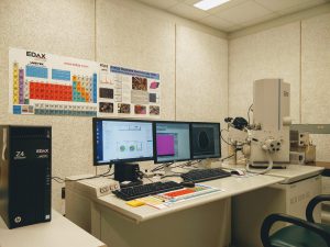
The Field-Emission Environmental SEM Philips XL30 is an instrument in which specimens can be examined at conventional high vacuum mode or at relatively high chamber pressure environment. It combines two main advantages:
- High resolution/magnification and excellent signal to noise ratio in both regular high vacuum and environmental mode due to field-emission electron source.
- Real “Wet” mode (100% humidity in the specimen chamber) and the possibility to examine specimens with pressure in the chamber up to 10 Torr.
Almost any specimen of suitable size can be examined in this microscope without destruction and additional specimen preparation procedures, which may lead to various artifacts.
Go here for specifications
Environmental Mode
By utilizing Peltier cooling stage and high water vapor pressure in the specimen chamber (so called “Wet” mode) it is possible to achieve high humidity (up to 100%) inside the chamber. In these conditions wet or hydrated specimens (cells, plant samples, tissue, etc.) will not dry and introduce any artifacts. Dynamic experiments are also possible; for example, drying or crystallization processes can be examined.
Specimen observation at raised chamber pressure without cooling stage (so called “variable pressure” mode) is useful for observing non-conductive dry specimens such as paper, plastics, ceramics, fibers, fabrics, outgassing materials.
The XL30 environmental microscope provides true secondary electron imaging, and so achieves very high resolution and can use low beam intensity. This is very important for beam-sensitive specimens such as biological samples, plastics, textile fibers.
High Vacuum Mode
The main advantage of the XL30 in high vacuum mode is its field-emission electron gun and advanced electron optics. A high resolution/magnification is obtainable at about any high voltage setting.
High vacuum mode can be divided in two sub modes:
- Low voltage mode – for observing non coated samples. Very low voltage (0.2-1.0 kV) can be used for observation of beam sensitive samples.
- Regular mode – for observing conductive and coated samples. Since useful resolution is very specimen dependable, this mode usually yields best results at highest magnifications.
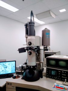
The CM12 STEM combines the features of both TEM and SEM. It can be used in standard TEM mode for thin samples or in scanning mode for thin or bulk samples. In scanning mode, a high brightness source (LaB6) produces a focused beam with high current density and small diameter for EDS X-ray microanalysis. Spatial resolution for microanalysis (a few nanometers for thin specimens) is much better than it is for microanalysis in SEM for bulk samples (about 0.5-3 microns). The STEM has a wide range of imaging and analytical capabilities, including a EDS system.
The CM12 STEM is equipped with the large format (11H Megapixel) retractable and fiber-optical coupled CCD camera SC100 ORIUS© CCD camera for digital image acquisition.
| High voltage range | 20 to 120 kV |
| Electron gun | LaB6 |
| Eucentric goniometer stage | ±60° |
| Camera length | 25 to 6500 mm |
| STEM magnifications | 70 to 510,000 |
| Minimum focused probe | 2 nm |
| Image media | Scanning mode – digital storage and/or Polaroid film. TEM mode – photo plates. |
| Standard, Be (for EDS) and Bulk side-entry specimen holders Digital EDS Prism detector with IMIX-PC system |
|