
Light Microscopy
Light Microscopy Core Facility
Director: Dr. Sarah Dallas
Other Personnel: Mark R. Dallas (Research Assistant, UMKC School of Dentistry)
Light microscopy underpins much of the research carried out within the mineralized tissue research programs at UMKC. Our microscopy core facility must continue to enable researchers to generate high quality photomicrographs of their various research applications.
The goals of the light microscopy core are to support mineralized tissue research within UMKC by:
- Providing high quality multi-user microscopes configured for brightfield and fluorescence microscopy and with high quality digital photography capabilities.
- Providing user training and support for the use of these microscopes.
- Continuing to make this service available to investigators within the Mineralized Tissue Center at no or minimal cost.
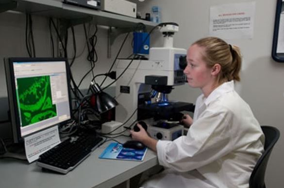
Support from the mineralized Tissue Center has enabled us to upgrade our Nikon E800 upright microscope with an automated x,y stage, fluorescence and DIC optics, high quality color and cooled CCD cameras and state of the art software. With the support of the Mineralized Tissue Center, we are also trying to leverage funding for additional state of the art light microscopy instrumentation, including a confocal microscope system with spectral unmixing capabilities and a multi-photon imaging system for live cell imaging.
Multi-User Microscopes Currently Available Within the Core:
Microscope #1- Nikon Eclipse E800 Upright Microscope
Configured for brightfield, phase contrast and fluorescence microscopy applications. The system has an Optronics cooled CCD color/black and white camera, interfaced with the AnalySIS software. Via an auxiliary Sony live video camera, the system can also be interfaced with the Osteomeasure software for quantitative bone histomorphometry.
Applications supported- This microscope system supports brightfield, phase contrast and fluorescence microscopy on specimens (e.g. tissue sections, cells) that are coverslip-mounted on microscope slides. The system also supports histomorphometric measurements of standard stained sections, fluorochrome labeled or immunofluorescently stained sections.
Microscope #2- Nikon Eclipse E800 Upright Microscope
Recently upgraded with support from the Mineralized Tissue Center, this microscope is configured for brightfield, DIC and fluorescence microscopy applications. The system has a Roper Scientific color CCD camera, a Photometrics low light monochrome CCD camera and a motorized x,y stage. The system is interfaced with the metamorph software for image acquisition and control of the hardware. Applications supported- This microscope system supports brightfield, DIC and fluorescence microscopy, including automated montage imaging on specimens (e.g. tissue sections, cells) that are coverslip-mounted on microscope slides.
Microscope #3- Nikon Eclipse TE300 Inverted Microscope
This inverted microscope system is configured for brightfield, phase contrast and fluorescence microscopy applications on specimens of varying thickness. The system has a Photometrics cooled CCD monochrome camera for fluorescence photography and a Roper Scientific color CCD camera for brightfield photography, interfaced with Q-imaging acquisition software. Applications supported- This inverted microscope system supports brightfield, phase contrast and fluorescence microscopy on samples of varying thickness. Its main use is for viewing live or fixed cells grown on various types of tissue culture vessels, such as culture plates and flasks.
Microscope #4- Nikon SMZ 445 Stereomicroscope
This microscope is configured for low power brightfield microscopy of larger three dimensional specimens, such as whole teeth, whole rodent bones and embryos. Illumination is via a fiber optic illumination system. This microscope has a Photometrics color/black and white digital camera, interfaced with the AnalySIS software. Applications supported- This microscope system supports low power microscopy and digital photography (i.e. “macro imaging”) of larger three dimensional samples, such as whole teeth, whole rodent bones and embryos. It is also used for magnifying specimens during microdissection.